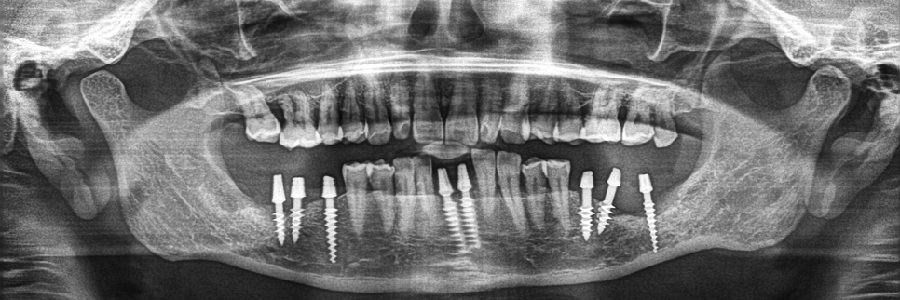


Modern Dentistry which has evolved rapidly in the past few years has brought the use of the latest technology to the world of dental sciences also, especially in Dental X-Ray OPG and Diagnostics. Dental X-Rays are pictures of the teeth, bones, and surrounding soft tissues to screen for and help identify problems with the teeth, mouth, and jaw. X-Ray pictures can show cavities, hidden dental structures, and bone loss that cannot be seen during a visual examination. Dental X-Rays may also be done as follow-up after dental treatments.
Oakwood Family Dental Clinic is well equipped with a state of the art minimum radiation. Digital X-Ray machine which allows minimum radiation exposure to the dental patient and the operator.
The following types of dental X-Rays are commonly used.
Bitewing X-Rays use the least amount of radiation and show the upper and lower back teeth in a single view. They are used to detect decay between the teeth and to show how well the upper and lower teeth line up. They also show bone loss that usually indicates the presence of severe gum disease or a dental infection.
Periapical X-Rays show the entire tooth, from the exposed crown to the end of the root and the bones that support the tooth. These X-Rays are used to detect dental problems below the gum line or in the jaw, including the presence of impacted teeth.
Occlusal X-Rays show the roof or floor of the mouth and are used to detect the presence of extra teeth, teeth that have not yet broken through the gums, jaw fractures, a cleft in the roof of the mouth (cleft palate), cysts, abscesses, or growths (such as a tumour). Occlusal X-Rays may also be used to locate foreign objects.
Panoramic Dental X-ray shows the entire mouth in single image, it is a two-dimensional (2-D) dental x-ray examination that captures the entire mouth. It doesn't produce the detail needed to show cavities.